Quantitative flow cytometry technology
On a small sample volume containing few cells and heterogeneous cell populations, flow cytometry allows to analyze with high sensitivity the cell size, its contents and the intensity of these stained cells.
Although in current practice of immunophenotyping flow cytometers are mainly used to differentiate positive and negative staining of cells, BioCytex uses these same instruments to quantitate molecules bound on cells, receptors, ligands or drugs as well as intracellular targets.
How does quantitative flow cytometry work ?
Quantitative flow cytometry is achieved by comparing fluorescence intensity and a calibration standard.
BioCytex uses a calibrator made of a mix of bead populations . These calibration beads are coated with known and increasing numbers of immunoglobulins (IgG) bound to their surface thus mimicking the binding of specific monoclonal antibodies to the biological sample cells.
Then, this calibrator and the target stained cells are incubated with polyclonal antibodies labeled with FITC fluorochrome.
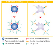
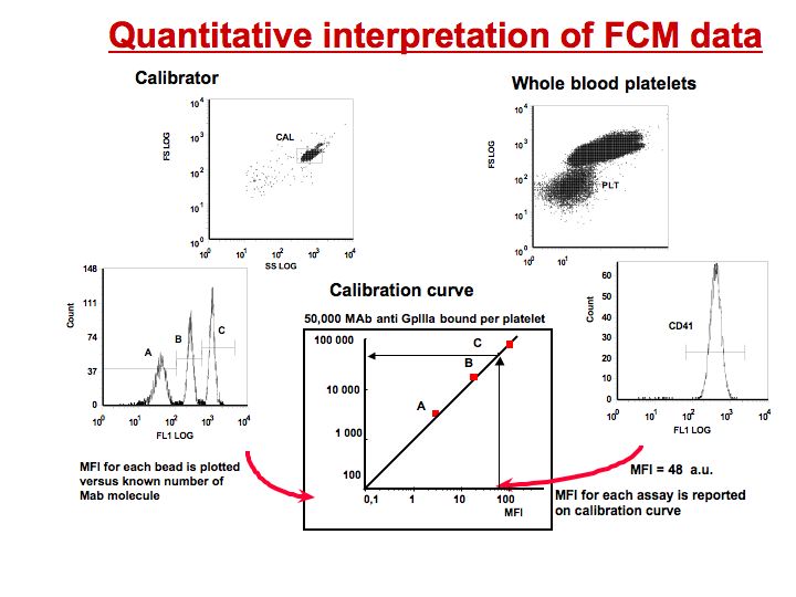
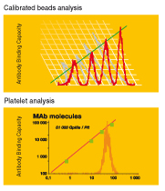
The beads bear specific number of sites and will allow to draw a calibration curve relating mean fluorescence intensity into a number of receptors per cell. The mean fluorescence intensity (MFI) of the tested sample is reported on the calibration curve to determine the number of molecules expressed per cell. Non specific staining is evaluated using an irrelevant antibody (negative isotypic control).
Quantitative interpretation of FCM data
The critical parameters to succeed in antigen quantitation include :
- Reaching saturation of antigen sites with the appropriate IgG monoclonal antibodies
- Using appropriate isotypic controls to evaluate specific binding, especially for low level detection
- Using beads which mimic cells bearing different amounts of antibody. The beads must offer specific features such as size and background fluorescence to correctly match the features of the cells of interest
- Calibrating the fluorescence response of the instrument
- Standardizing and calibrating the antibody binding capacity. Even in the same cluster differentiation (CD) specificity, several monoclonal antibodies may reveal different numbers of antigen sites since they will recognize different epitopes
- Finally, pre-analytical variables have to be established and controlled for any specific application.
These requirements has led BioCytex to the development of kits including all necessary reagents, not only calibrators but also antibodies, buffers, agonists and a written procedure for in vitro diagnostic use of these applications.
The Biocytex product line can be categorized as follows:
- Drug responsiveness (PLT VASP/P2Y12, PLATELET GpIIb/IIIa Occupancy)
- Thrombotic risk (CELLQUANT PNH, REDQUANT PNH, PLT VASP/P2Y12, PLATELET GpIIb/IIIa Occupancy)
- Bleeding disorders (PLT HPA-1, PLT Gp/Receptors, PLATELET PAIg, PLATELET Gp Screen, PLT VASP/P2Y12)
- Vascular biology (CY-QUANT ELISA sCD146, Megamix)
- Customizable kits (PLATELET Calibrator, CELLQUANT Calibrator)
Why using Biocytex quantitative flow cytometry technology ?
Numerous biological molecules present an interest in the measurement of their expression level. Indeed any modulation of receptor expression level associated to physiological differentiation, pathological state or therapeutic treatment brings valuable information to the biologists and the physicians.
Such measurements in Clinical Biology require full reliability in term of day to day, inter instrument and lab-to-lab reproducibility.
As an example, an ambulatory patient treated with a new generation of antiplatelet drugs will be easily monitored wherever he travels.